


 中 文
中 文
Leading Countries in the Global Dental Industry: Companies and Advanced Technologies
2025-03-22
2026-09-29
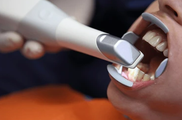
Intraoral scanning is often described as an optical process, but the final digital impression is not simply a direct "picture" of the patient's teeth.
The scanner first captures optical information through its imaging system. That raw information then goes through calibration, geometric correction, three-dimensional reconstruction, frame registration, stitching, and other software processing before becoming the final digital model.
This raises an important question:
If software corrects or compensates for errors in the scanning process, how do we know that the correction itself is correct?
The answer is not that the software is assumed to be accurate. Instead, the accuracy of the complete scanning system is evaluated by comparing its output with a reliable reference.
An intraoral scanner needs to convert the physical surface of teeth and oral structures into digital three-dimensional data.
A simplified workflow can be described as:
Optical acquisition → Calibration → 3D reconstruction → Registration → Stitching → Software processing → Digital model
Each stage can influence the final result.
The optical system may introduce errors related to factors such as lens characteristics, sensor performance, optical distortion, and measurement geometry.
The software then processes the captured information and reconstructs the three-dimensional surface.
Therefore, the accuracy of an intraoral scanner should not be understood as the accuracy of its optical components alone.
What matters clinically is the accuracy of the final digital impression.
Software correction is not simply a matter of adding or subtracting a fixed value from every measurement.
In a real scanning system, different sources of error can have different characteristics.
For example, errors may be related to:
During system development, known reference objects can be used to characterize the behavior of the scanning system.
The resulting calibration and correction models can then be incorporated into the software.
In simplified terms:
Known reference → Measured data → Difference identified → Correction model → Corrected output
However, identifying a correction model does not automatically prove that the final output is accurate.
The correction must still be validated against reference data.
This is one of the most important questions in evaluating digital scanning accuracy.
The software cannot prove its own accuracy.
Instead, the final digital result is compared with an independent or established reference.
A simplified validation process is:
Reference object
↓
High-accuracy reference measurement
↓
Reference 3D dataset
↓
Intraoral scanner
↓
Digital scan
↓
3D comparison
↓
Deviation analysis
If the digital scan is sufficiently close to the reference dataset under defined test conditions, the complete scanning workflow can be considered to have achieved the corresponding level of accuracy.
This means that the evaluation is not simply asking:
"Is the software correction correct?"
It is asking:
"After optical acquisition, calibration, software processing, and correction, how close is the final digital model to the reference?"
This distinction is important.
A reference dataset provides a basis for evaluating the scanner's output.
For example, a test object can first be measured using a high-accuracy reference measurement system. The resulting dataset represents the geometry against which the intraoral scanner output can be compared.
The same object is then scanned with the intraoral scanner.
The two three-dimensional datasets are aligned and compared.
Conceptually:
Reference 3D dataset
vs.
Intraoral scanner 3D dataset
↓
Surface deviation
The difference between the two datasets provides information about how closely the scanner reproduced the reference geometry.
Research evaluating intraoral scanners commonly follows this general approach. For example, studies have compared repeated intraoral scans with reference datasets obtained using high-precision scanning systems and then analyzed the deviation between the datasets.
When discussing scanning accuracy, two concepts are particularly important:
Trueness describes how close the average result is to the accepted reference value.
In practical terms:
Does the digital model reproduce the actual geometry correctly?
Precision describes how close repeated measurements are to one another.
In practical terms:
If the same object is scanned multiple times, do the results remain consistent?
These are related but different characteristics.
A scanner could produce highly repeatable scans that are consistently shifted from the reference. In that case, precision may be good while trueness is poorer.
Conversely, a scanner may produce results that are close to the reference on average but show greater variation between repeated scans.
Therefore, evaluating only one number is not always sufficient to describe the complete measurement performance.
The basic principle is straightforward:
Reference geometry → Scan geometry → Spatial comparison → Deviation
However, the actual analysis can be more sophisticated than measuring the distance between two individual points.
A complete three-dimensional dataset contains a large number of surface points.
After the datasets are appropriately aligned, the software can calculate the spatial deviation between corresponding surface regions.
The resulting data can then be summarized using statistical measures such as:
A color deviation map can also be generated to show where the scanned surface differs from the reference.
This is why a statement such as:
"Accuracy: 20 μm"
should not be interpreted without knowing how that value was obtained.
The number could represent a particular statistical measurement under a specific test condition rather than the maximum error at every point on the scanned surface.
Yes.
ISO 20896-1:2019 is specifically dedicated to methods for assessing the accuracy of digital impression devices using handheld scanning devices to acquire three-dimensional descriptions of intraoral surfaces. ISO states that the standard specifies test methods and procedures for this purpose, and the 2019 edition was reviewed and confirmed in 2025, meaning it remains current.
Importantly, the standard is not simply a formula for calculating one "accuracy" number.
It provides a broader testing framework covering areas such as:
The standard also includes different test-object configurations, including crown preparation, inlay preparation, and full-arch testing.
Therefore, it is more accurate to say:
ISO 20896-1 provides standardized test methods and procedures for assessing the accuracy of digital impression devices.
It would be less accurate to say that the standard simply defines one universal formula that every intraoral scanner must use to calculate an "accuracy" value.
Even when two scanners report accuracy values in micrometers, those values should not automatically be treated as directly comparable.
The testing conditions matter.
For example:
| Factor | Why It Matters |
|---|---|
| Reference measurement | Determines the basis of comparison |
| Test object | Different geometries create different challenges |
| Scanning area | Full-arch scanning introduces different registration challenges |
| Number of repeated scans | Influences precision evaluation |
| Alignment method | Affects the calculated deviation |
| Statistical metric | Different metrics describe different aspects of error |
| Software version | Algorithms can influence the final result |
ISO 20896-1 itself includes requirements concerning reference measurement, test objects, test conditions, digital impression processing, accuracy assessment, and test reporting.
Therefore, comparing two isolated numbers without knowing their testing conditions can be misleading.
Yes.
Software is not merely a user interface placed on top of the optical hardware. It can influence how captured data are reconstructed, registered, and ultimately converted into the final digital impression.
Research has demonstrated that software updates can change the trueness and precision of intraoral scanners.
In one study, different software versions produced statistically significant differences in scanning accuracy. The effects were not always in the same direction, and the influence also varied according to the scanned region and material.
Another study evaluated software updates by repeatedly scanning reference models and comparing the resulting datasets with high-precision reference scans. The researchers found that software version could affect both trueness and precision.
This leads to an important conclusion:
Software is part of the measurement system.
A change in software can potentially change the characteristics of the final digital impression, even when the physical scanner hardware remains unchanged.
Not necessarily.
It may seem logical that a newer software version should always produce a more accurate result.
However, experimental evidence shows that the relationship is not necessarily linear.
Different software versions can affect different scanning regions differently, and newer software does not automatically guarantee better performance in every test condition.
This is why accuracy needs to be demonstrated through testing rather than assumed from the software version number.
In other words:
Newer software ≠ automatically more accurate
Instead:
New software → needs validation → measured performance
Single-tooth scanning and full-arch scanning should not be treated as exactly the same measurement problem.
When scanning a single preparation, the scanner only needs to reconstruct a relatively limited area.
During full-arch scanning, the system continuously captures and registers multiple frames.
A simplified process is:
Frame 1 → Frame 2 → Frame 3 → Frame 4 → … → Frame N
Each frame needs to be registered with the previous data.
Small registration errors can accumulate as the scanning distance increases.
Therefore, a scanner can perform very well on a small scanning area while showing different behavior during full-arch scanning.
This is one reason why accuracy data should always be considered together with the scanning range and test configuration.
Standardized testing provides a controlled way to evaluate scanning performance, but clinical scanning introduces additional variables.
In a laboratory test, the object may have:
The oral environment is much more complex.
Operator experience, scanning strategy, patient anatomy, moisture, surface characteristics, and scanning conditions can all influence the resulting digital impression. Research has also shown that operator experience and training can influence scanning accuracy.
This means that a laboratory accuracy value should not automatically be interpreted as the exact error that will occur in every clinical case.
When evaluating an intraoral scanner, it is useful to ask more than:
"What is the accuracy?"
A better set of questions is:
These questions help turn a marketing specification into a measurable technical claim.
The accuracy of an intraoral scanner is therefore not determined by its optical system or software correction alone.
A more complete picture is:
Optical acquisition
↓
System calibration
↓
Geometric and optical correction
↓
3D reconstruction
↓
Frame registration and stitching
↓
Software processing
↓
Final digital impression
↓
Comparison with reference data
↓
Deviation analysis
↓
Trueness + Precision
This entire chain matters.
The software does not need to be "perfect" in isolation. What matters is whether the complete measurement system produces a digital model that is sufficiently close to the reference under defined conditions.
Optical components and software correction are both fundamental to the accuracy of intraoral scanning, but neither should be evaluated in isolation.
The optical system captures the information. Calibration and software processing compensate for known system characteristics and transform the captured information into a three-dimensional digital model. The accuracy of the final result is then evaluated against a reference rather than simply assumed to be correct.
ISO 20896-1:2019 provides a standardized framework for assessing the accuracy of digital impression devices using handheld scanning devices. It addresses reference measurements, test objects, test conditions, digital impression processing, accuracy assessment, and test reporting.

Dry & wet milling for zirconia, PMMA, wax with auto tool changer.
learn more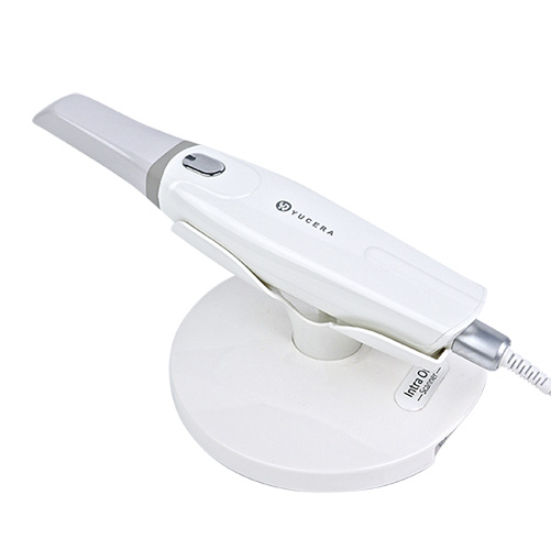
High-precision 3D scanning, AI calibration, full-arch accuracy.
learn more
40-min full sintering with 57% incisal translucency and 1050 MPa strength.
learn more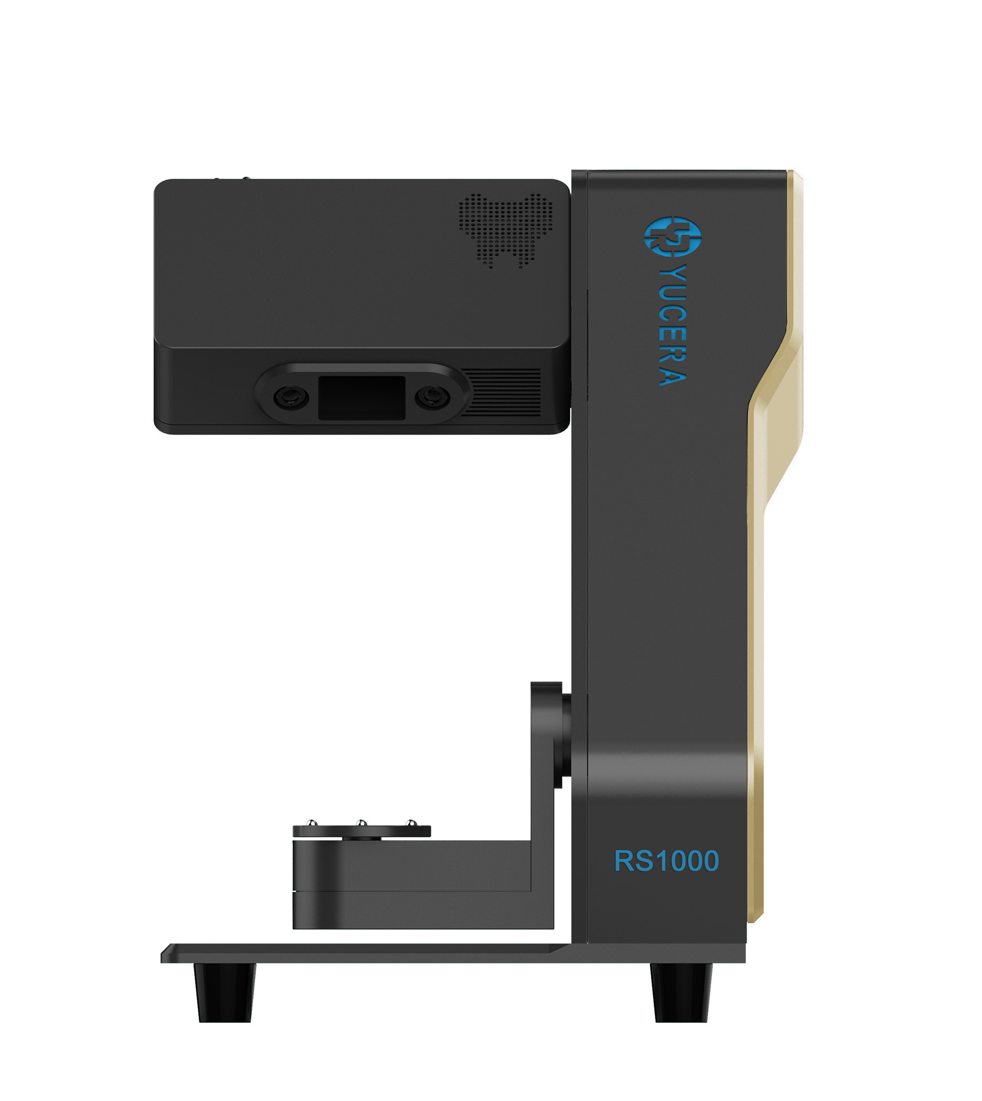
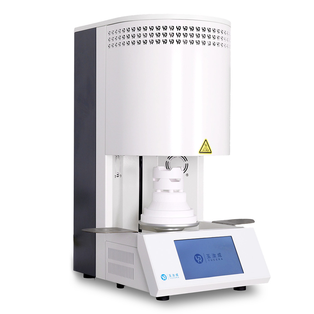
40-min cycle for 60 crowns, dual-layer crucible and 200°C/min heating.
learn more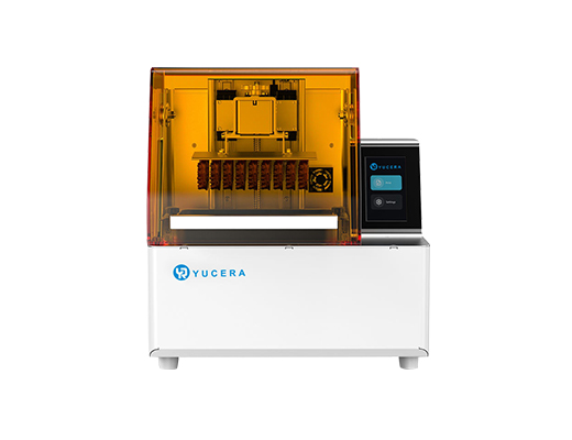
High-speed LCD printer for guides, temporaries, models with 8K resolution.
learn more


2025-03-22
2026-06-05

2025-04-02


